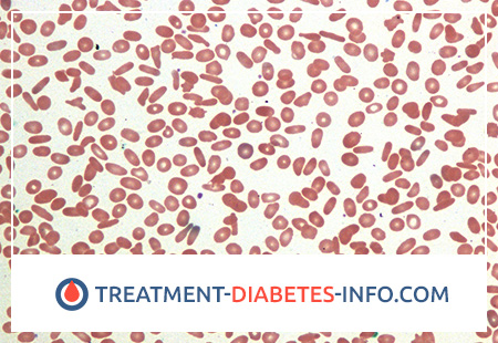
What is Hereditary Elliptocytosis?
Hereditary elliptocytosis (synonym – ovalocytosis) is a hereditary deterministic disease transmitted by an autosomal dominant mode of inheritance. The erythrocyte anomaly that develops is characterized by a disorder of the structure of the erythrocyte membrane.
Hereditary elliptocytosis, or oval-cell anemia, is a hereditary form of hemolytic anemia. Oval erythrocytes (elliptocytes) are observed in healthy people, their number can reach 10%. With hereditary elliptocytosis, oval erythrocytes are significantly larger, in the range of 25-75%.
Hereditary elliptocytosis is usually divided into three large groups:
- hereditary elliptocytosis with discoidal elliptocytes;
- spherocytic or ovalocytic hereditary elliptocytosis;
- hereditary elliptocytosis with the presence of stomatocytes (also called Melanesian or South Asian ovalocytosis), in which erythrocytes have a rounded shape with an unpainted area in the center of the erythrocyte, bounded by two curved lines connected to the sides.
Causes of Hereditary Elliptocytosis
The frequency of the elliptocytosis gene in the human population is 0.02-0.04%, the disease occurs with the same prevalence rate as microspherocytosis, but is less often diagnosed, since a significant proportion of carriers lack any manifestations of the disease. During electrophoresis, in some patients there is no protein band 4.1. Red blood cells have the shape of an ellipse, their deformability is reduced, and therefore they are quickly destroyed in the spleen.
Pathogenesis during Hereditary Elliptocytosis
The morphological examination of the blood makes it possible to make a diagnosis, but often, along with elliptocytosis, a certain amount of microspherocytes is detected in a smear, poikilocytosis is detected. With elliptocytic hemolytic anemia, the osmotic resistance of erythrocytes is significantly reduced, as well as increased erythrocyte self-destruction, which is completely normalized by glucose.
Deficiency of glycophorin C may occur due to individual molecular defects. In individuals homozygous for glycophorin C, an elliptocytosis without anemia is observed.
Erythrocyte progenitors with hereditary elliptocytosis have a rounded shape: erythrocytes acquire an elliptoid form only when leaving the CM. It is assumed that red blood cells with hereditary elliptocytosis are constantly stabilized on their pathological form by reorganization of the skeleton when cells come into contact with new proteins (blood serum).
Symptoms of Hereditary Elliptocytosis
In most cases, this anomaly does not give any clinical manifestations, however, in some patients, the disease is accompanied by hemolytic anemia with intracellular disintegration of erythrocytes, mainly in the spleen.
The normal content of oval erythrocytes in healthy people can reach 10%. In patients with hereditary elliptocytosis, elliptocytes make up 25-75%.
The literature describes many cases of a combination of elliptocytosis with other forms of hereditary anemia, such as sickle cell anemia, thalassemia.
An asymptomatic form of elliptocytosis, elliptocytic hemolytic anemia as a nosological form, and symptomatic elliptocytosis in other diseases are distinguished.
The course of hereditary elliptocytosis is overwhelmingly (95%) asymptomatic. However, it should be remembered that the presence of elliptocytosis does not always indicate its hereditary character, since in a healthy person about 15% of erythrocytes also have an ellipsoid shape. Clinically in manifest cases, yellowness of the skin and sclera, splenomegaly are observed, cholelithiasis, changes in the bone skeleton are often diagnosed.
Diagnosis of Hereditary Elliptocytosis
The differential diagnosis of the disease is carried out with symptomatic elliptocytosis, possible with subleukemic myelosis. In this case, the patient is carried out a study of histomorphology and bone marrow architectonics. It is very important to examine the relatives of the patients. Ovoid erythrocytes can be detected and vitamin-B12-deficient anemia.
Laboratory diagnosis of hereditary elliptocytosis is based on the identification of elliptocytes, which sometimes have the shape of a rod. If normally the ratio of mutually perpendicular diameters of an erythrocyte approaches 1, then with elliptocytosis it decreases to 0.78. Target red blood cells can occur, and elliptocytes can be of different sizes and normochromic coloration.
The color indicator does not deviate from the norm, the hemoglobin level even in homozygotes is not low, varying from 90 to 120 g / l. The number of reticulocytes increases moderately – up to 4%; osmotic resistance of erythrocytes is often reduced, but may be normal; in the latter case, samples are performed with incubation of erythrocytes and a sample for autolysis, which show a decrease in the osmotic resistance of erythrocytes.
Treatment of Hereditary Elliptocytosis
No treatment is required for abnormalities; in elliptocytic hemolytic anemia, removal of the spleen is as effective as in microspherocytosis.
Therapeutic measures for elliptocytic hemolytic anemia are similar to those used for hereditary microspherocytosis.
The prognosis is favorable in patients undergoing splenectomy, the rest may develop severe hemolytic crises, cholelithiasis, and thrombosis.
Prevention of Hereditary Elliptocytosis
Prevention of hereditary elliptocytosis is not carried out.

